
What is the mitral valve?
The mitral valve lies between the left atrium and left ventricle. It helps blood flow forward and prevents it from leaking backwards.
Mitral valve diseases
Common conditions include mitral regurgitation (leakage), mitral stenosis (narrowing) and mitral valve prolapse.
For information about their causes, symptoms and treatment, see the Mitral Valve Disease Guide for further details.
What causes mitral valve disease?
Mitral valve disease can have several causes:
- Rheumatic fever: An important cause of mitral stenosis, with its frequency varying between populations
- Degenerative changes: Changes in the valve tissue that may develop with age or occur earlier in life
- Mitral valve prolapse: One or both leaflets bulge back towards the left atrium
- Infection (endocarditis): Infection can damage the valve
- Damage to the papillary muscles after a heart attack
- Congenital valve abnormalities
- Connective tissue disorders, such as Marfan syndrome
When is surgery needed?
The decision depends on the severity of valve disease, symptoms, heart size and function, and other findings. In selected patients, intervention may be advisable before symptoms develop; a specialist valve team assesses the timing.
Surgical treatment options
What is mitral valve repair?
Repair corrects the valve problem while preserving the patient's own valve wherever possible, rather than replacing it with a prosthesis.
For information about suitability, techniques and potential benefits, see the Mitral Valve Repair Guide for further details.
Which techniques are used?
The surgeon may combine techniques according to the valve abnormality:
- Supporting the valve ring (annuloplasty): A ring or band reshapes and supports the valve opening to help the leaflets meet
- Reshaping excess valve tissue: Selected areas of excess or prolapsing tissue may be removed or reshaped
- Repairing the supporting cords (chordae): Stretched or torn cords may be repaired or replaced with artificial cords
- Helping the leaflets meet: The leaflets can be supported or brought together to improve closure
- Treating fused or calcified areas: Selected areas may be treated when appropriate; extensive calcification can limit the possibility of repair
In conditions with excessive leaflet movement, such as Barlow's disease, repair may be possible in experienced centres, but suitability and durability require individual assessment. Read more in Barlow's Disease and Mitral Valve Repair for additional information.
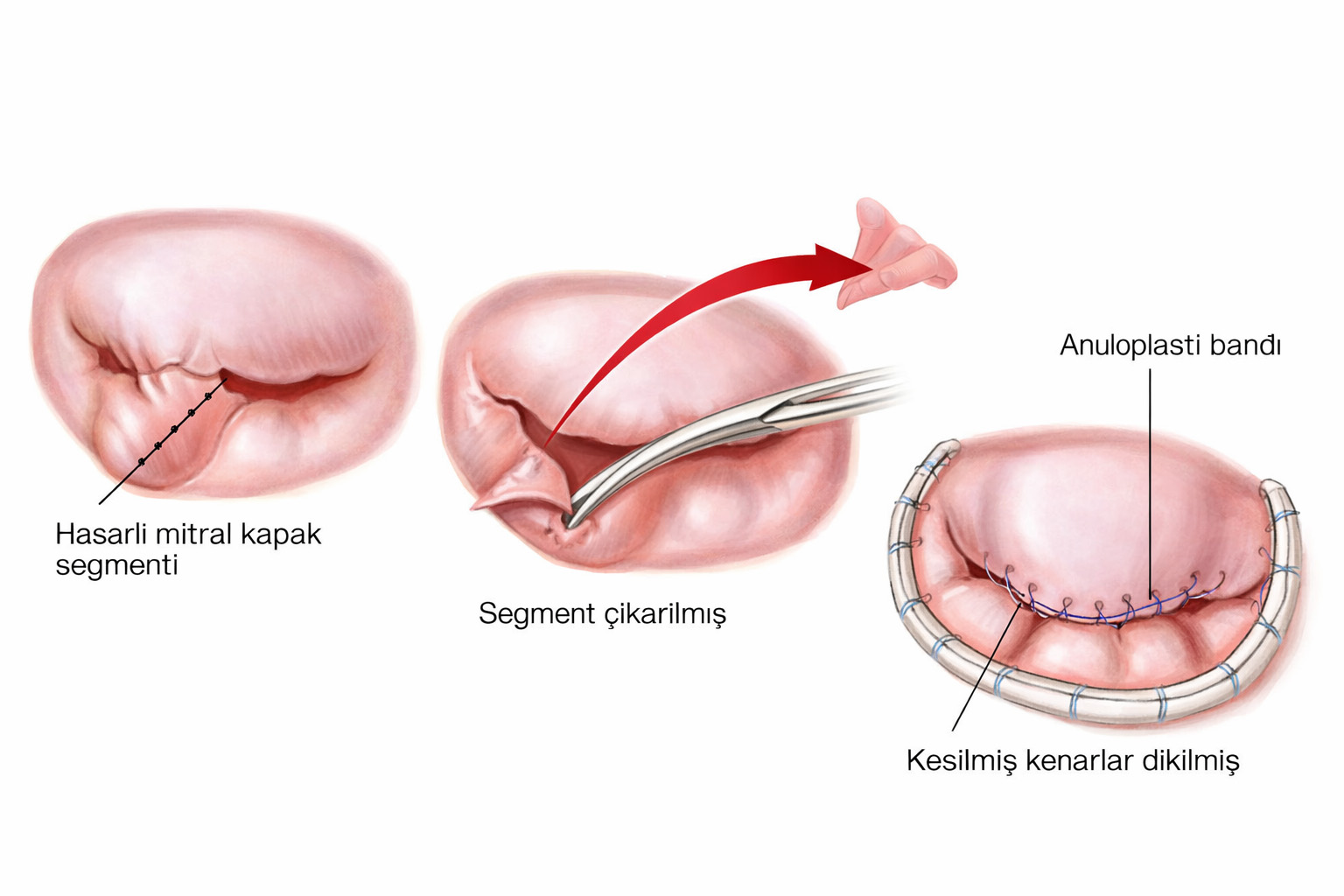
Potential advantages of mitral valve repair
What is mitral valve replacement?
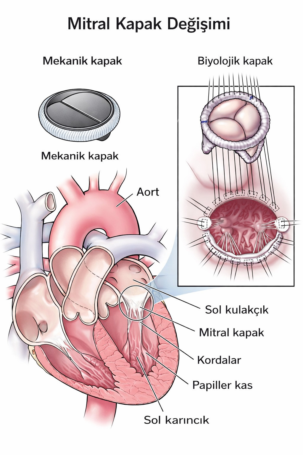
Replacement places a prosthetic valve in the position of the diseased valve. It may be necessary for severe narrowing or leakage when a satisfactory, durable repair is not feasible.
Types of replacement valve
⚙️ Mechanical valve
- Very durable, although no prosthesis has an unlimited guarantee
- Requires lifelong treatment with a vitamin K antagonist, such as warfarin, and regular monitoring
🫀 Biological (tissue) valve
- Usually made from animal tissue
- Does not necessarily require lifelong anticoagulation solely because of the valve, but short-term treatment or another medical indication may still require it
- Can deteriorate over time and may eventually require another intervention
Catheter-based treatment options
Selected patients, including some with a high surgical risk, may be suitable for catheter treatment. Suitability depends on the cause of leakage, valve anatomy and specialist Heart Team assessment.
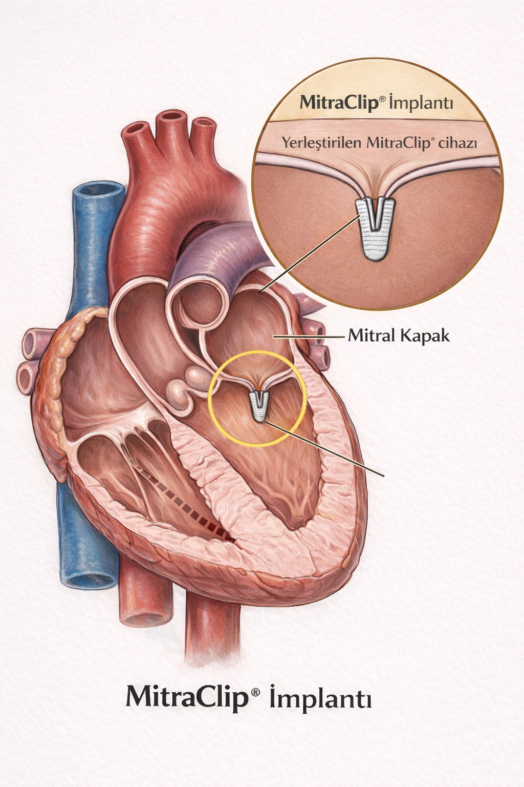
MitraClip
A catheter introduced through a vein in the groin delivers a device that brings parts of the mitral leaflets together. This transcatheter edge-to-edge repair does not require open-heart surgery and is appropriate only for selected patients.
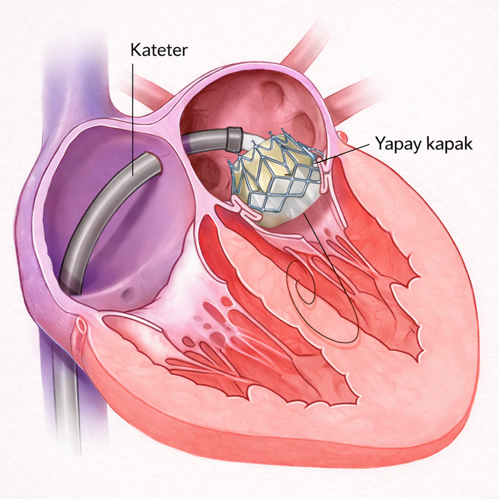
Transcatheter valve implantation
Catheter-based mitral valve implantation is an option in selected situations, such as some failing tissue valves. Native-valve replacement has specific anatomical and device requirements and is assessed in specialist centres.
Surgical approaches
Mitral valve surgery generally uses a heart-lung machine and usually involves temporarily stopping the heart. The surgical approach is selected according to the patient's anatomy and condition.
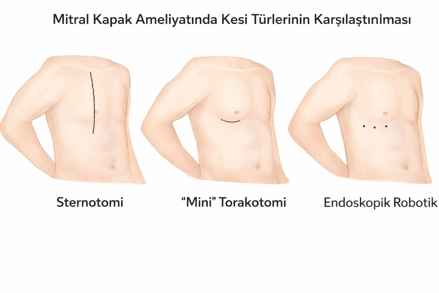
- Conventional surgery: Usually performed through a midline incision in the chest and division of the breastbone.
- Minimally invasive surgery: Uses smaller incisions, often on the right side of the chest, with additional access in the groin when needed. It remains heart surgery and is not the same as catheter treatment.
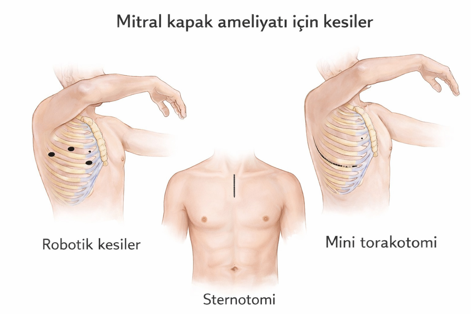
Minimally invasive surgery may use these techniques:
- Endoscopic surgery: The surgeon operates with long instruments while viewing the heart through a camera introduced into the chest.
- Robotic surgery: The surgeon controls robotic instruments from a console while viewing the operative field through a camera. The robot does not perform the operation independently.
Recovery after surgery
For further information about recovery, see Mitral Valve Disease — Frequently Asked Questions for additional information.
- Initial monitoring in intensive care, often for about one day but longer if needed
- Several further days in hospital; the length of stay depends on recovery and complications
- Activity increases gradually over several weeks; full recovery after open surgery may take around two to three months or longer
Key point
Mitral valve surgery can improve symptoms and quality of life. Benefits, risks and recovery vary with the underlying disease, heart function and the procedure performed.


