
Contents
The Normal Aortic Valve and Its Physiology
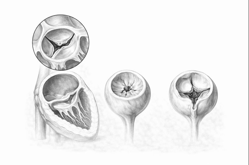
A normal aortic valve usually consists of three thin, flexible leaflets, or cusps. During systole, the contraction phase of each heartbeat, they open fully to let blood flow from the left ventricle into the aorta. During diastole, the relaxation phase, they close tightly to prevent blood from leaking back into the heart.
Aortic Stenosis and Regurgitation: Key Differences
| Feature | Aortic Stenosis | Aortic Regurgitation |
|---|---|---|
| Definition | A narrowed valve opening causes pressure overload | Incomplete valve closure causes volume overload |
| Common Causes | Age-related calcification, bicuspid valve, rheumatic disease | Aortic root dilation, bicuspid valve, connective tissue disorders |
| Typical Symptoms | Breathlessness, chest pain (angina), fainting (syncope) | Breathlessness, fatigue, palpitations, heart enlargement |
| Treatment | Surgical AVR; TAVI in suitable patients | Repair if feasible; otherwise replacement |
Symptoms and Their Underlying Mechanisms
- Breathlessness and Reduced Exercise Capacity: These are early signs of heart failure in both conditions.
- Chest Pain (Angina): Particularly typical of aortic stenosis. The increased oxygen demand of the thickened heart muscle cannot be met.
- Fainting (Syncope): One of the most serious symptoms of aortic stenosis. Blood flow to the brain decreases during exertion.
- Palpitations: Especially in aortic regurgitation, stronger heartbeats are felt as the heart pumps an increased volume of blood.
- A Murmur on Examination: Often the first finding: an abnormal sound made by blood flowing through the diseased valve.
Causes and Risk Factors
- Age-Related Calcification: This is the most common cause of aortic stenosis, particularly in people over 70.
- Bicuspid Aortic Valve: A congenital condition in which the valve has two leaflets instead of three. Abnormal blood flow causes earlier calcification, often between ages 40 and 60.
- A History of Rheumatic Fever: It can cause adhesions and thickening of the valve leaflets.
- Aortic Root Dilation (Aneurysm): It stretches the valve annulus, preventing the leaflets from meeting and causing regurgitation.
Diagnosis: The Role of Echocardiography
Although hearing a murmur on examination starts the diagnostic process, for a definitive assessment transthoracic echocardiography is the gold standard. Echocardiography assesses valve structure, stenosis severity, aortic valve area (AVA) and the heart’s pumping function, or ejection fraction (EF). An AVA below 1 cm² is considered severe stenosis.
Treatment Options
Medication
Symptom control in mild or moderate disease. It cannot correct the mechanical defect.
Valve Replacement (AVR)
A definitive solution for severe valve disease. A mechanical or biological valve is selected.
TAVI
A new biological valve is inserted by catheter through the groin. In suitable patients, the chest is not opened.
Minimally Invasive / Sutureless
Mini-sternotomy or right thoracotomy. Less pain and faster recovery.
Surgical Aortic Valve Replacement (AVR)
⚙️ Mechanical Valve
Highly durable and designed to last a lifetime. However, it requires lifelong anticoagulation with warfarin and regular INR monitoring. It is preferred in younger patients.
🫀 Biological Valve
It does not require lifelong anticoagulation after the first three months. However, it may deteriorate over 10–20 years and require another intervention. It is preferred in older patients.
TAVI (Transcatheter Aortic Valve Implantation)
Instead of open-heart surgery, a catheter is introduced through an artery in the groin to place a new biological valve inside the diseased, calcified valve. Recovery is much faster because the chest is not opened. Initially used only in high-risk patients, it is now also considered in intermediate- and low-risk patients.
The Critical Factor: Timing
In severe aortic stenosis, average life expectancy falls to 2–3 years after symptoms such as breathlessness, chest pain and fainting develop. Delayed treatment can result in irreversible heart muscle damage.
Recovery After Surgery
Frequently Asked Questions
To Book an Appointment
Prof. Dr. Selim İsbir — Yeditepe University Koşuyolu Hospital


