
In this article, you can find all the details, from the causes of aortic arch aneurysm and its interesting symptoms like hoarseness, to open surgery, and modern Hybrid and Frozen Elephant Trunk methods.
Vital Junction: What is the Aortic Arch?
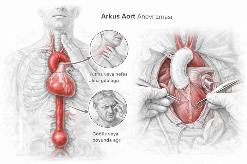
The aorta exits the heart upwards (Ascending Aorta), forms an arch at the base of the neck (Aortic Arch), and descends down the back (Descending Aorta). The arch region, or “bend,” is the body’s most strategic junction because three main vessels branch off from it:
[Image of Aortic Arch Anatomy]
- Brachiocephalic Artery: Carries blood to the right arm and the right side of the brain.
- Left Carotid Artery: The main carotid artery supplying the left side of the brain.
- Left Subclavian Artery: Carries blood to the left arm.
An aneurysm (dilation) in this region not only carries the risk of rupture; it can also directly threaten brain functions through clot formation or compression.
Why Does It Occur? Risk Factors
Factors that lead to weakening of the vessel wall include:
- Atherosclerosis (Hardening of the Arteries): Plaque buildup in the vessel wall due to advanced age, smoking, and high cholesterol damages the structure.
- Chronic Hypertension: Uncontrolled high blood pressure constantly impacts the aortic wall, causing it to dilate.
- History of Aortic Dissection (Tear): In patients who have previously experienced Type A dissection, dilation (residual dissection aneurysm) may occur over time in the segment beyond the surgically repaired area.
- Genetic Diseases: Connective tissue diseases such as Marfan Syndrome, Loeys-Dietz Syndrome.
Hoarseness and Other Symptoms
Aneurysms usually grow “silently.” However, when their diameter exceeds 5-6 cm, they can cause the following symptoms due to compression of surrounding tissues:
- Hoarseness: A very typical symptom. It occurs due to compression of the nerve (Recurrent Laryngeal Nerve) that passes directly beneath the aorta and goes to the vocal cords.
- Difficulty Swallowing: Occurs due to pressure on the esophagus, which is directly behind the aorta.
- Shortness of Breath and Cough: Seen as a result of compression on the trachea (windpipe).
- Chest and Back Pain: Usually a dull, deep, and throbbing pain. If it is sudden and tearing, it is a sign of an emergency.
Diagnostic Methods
Although diagnosis is often made incidentally, advanced imaging is essential for definitive evaluation and surgical planning.
- CT Angiography (Tomography): The gold standard. The aneurysm’s diameter, its relationship with brain vessels, and calcification status are seen in millimeters.
- MR Angiography: Does not involve radiation, preferred for patients requiring frequent follow-up or those with kidney failure.
- Echocardiography and TEE: Used to evaluate the condition of heart valves.
Treatment Options
For aortic arch aneurysms, medication is only for controlling blood pressure and slowing growth. Definitive treatment is surgical.
When is Surgery Decided?
- When the aortic diameter exceeds 5.5 cm.
- In genetic diseases like Marfan, the limit is 4.5 – 5.0 cm.
- Regardless of the diameter; if there is rapid growth or if compression symptoms (hoarseness, etc.) have appeared, surgery is essential.
1. Open Surgery (Total Arch Replacement)
This surgery is one of the most complex procedures in cardiac surgery. The breastbone is opened, and a heart-lung machine is used.
Special Technique: During the suturing of the parts of the vessels leading to the brain, body temperature is lowered to 20-25 degrees (hypothermia), and circulation in the lower body is stopped. During this time, blood continues to be supplied to the brain using special methods (selective cerebral perfusion). The goal is to protect the brain. The dilated vessel is removed and replaced with an artificial vessel (graft).
2. Hybrid Methods and the Elephant Trunk Technique
With technological advancements, “Hybrid” methods are gaining prominence.
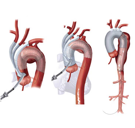
- Frozen Elephant Trunk: While the aortic arch is being replaced during open surgery, a stented artificial vessel extending into the descending aorta is also placed in the same session. This allows for the resolution of both anterior and posterior aortic problems in a single session.
- Debranching + TEVAR: In high-risk patients, brain vessels are bypassed and repositioned through small incisions in the neck region. Then, a stent (TEVAR) is inserted into the arch region via the groin. This provides treatment without fully opening the chest cavity.
Post-Surgery Recovery Process
The process after aortic arch surgery is slightly more delicate than standard heart surgeries.
- Intensive Care: Lasts an average of 2-3 days. During this period, awakening and neurological control (monitoring for stroke risk) are the most critical stages.
- Ward Follow-up: Lasts approximately 5-7 days. Hoarseness and swallowing functions are checked.
- Full Recovery: Bone healing and reaching full effort capacity takes 6-8 weeks.
Frequently Asked Questions (FAQ)
Is aortic arch surgery very risky?
These surgeries are “major” procedures requiring high technical skill. The most significant risks are bleeding and neurological events (stroke). However, in experienced centers and with modern brain protection techniques (cerebral perfusion), success rates are at 90-95%.
Will my voice be hoarse after surgery?
The nerves in the arch region (recurrent laryngeal nerve) are very close to the surgical field. Temporary hoarseness may occur, which usually resolves over time. The risk of permanent damage is low but present.
Can I be treated with a closed method (Stent)?
Since the arch region is the branching point for brain vessels, standard stents cannot be placed here (they would block brain vessels). However, with “Hybrid” methods or special fenestrated stents, closed or semi-open treatment is possible if the patient’s anatomy is suitable.
What happens if an aneurysm ruptures?
Aneurysm rupture is a life-threatening emergency that can lead to fatalities before reaching the hospital. Therefore, undergoing surgery under planned conditions, before the aneurysm ruptures, is the safest approach.
Conclusion and Recommendations
Aortic arch aneurysm, due to its location, carries vital risks and is one of the most challenging surgical areas. Early diagnosis and the correct treatment method are the most important factors determining life expectancy and quality. Thanks to developing hybrid surgical techniques, it is possible to achieve successful results with less invasive procedures. It is life-saving for individuals with risk factors to not neglect regular cardiological check-ups and to consult a specialist cardiovascular surgeon without delay if symptoms appear.