
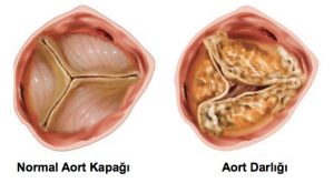
The Difference Between a Normal Aortic Valve and Aortic Valve Stenosis
What is Aortic Valve Stenosis?
Aortic valve stenosis is a condition where the valve between the main artery leaving the heart (aorta) and the left ventricle narrows. This narrowing prevents the heart from pumping enough blood to the body. As a result, oxygenated blood cannot be distributed sufficiently to meet the body’s needs, leading to various health problems.
What Are the Symptoms?
The disease may not show symptoms until severe movement restriction occurs in the valve. However, in later stages, the following symptoms are commonly seen:
- Shortness of breath
- Chest pain and pressure
- Fainting
- Heart palpitations
- Fatigue upon exertion
If these complaints are present, a cardiologist should be consulted immediately.
What Causes Aortic Valve Stenosis?
The most common cause is age-related calcium buildup. Calcification on the valve gradually narrows the valve opening. Additionally, people born with a bicuspid aortic valve (two leaflets instead of three) may develop this disease at an earlier age. In rare cases, rheumatic heart diseases can also lead to aortic valve stenosis.
Who is at Risk?
- Individuals over 60 years of age
- Those with congenital heart disease
- Those with a history of rheumatic disease
- The risk is slightly higher in men
In childhood, congenital aortic valve anomalies can lead to early diagnosis of the disease.
How is it Diagnosed?
Aortic valve stenosis is usually detected by hearing a heart murmur during physical examination. The definitive diagnosis is made with Echocardiography (ECO). This method examines the movement of heart valves, their openings, and blood flow in detail.
Treatment Methods
Unfortunately, there is no definitive medical treatment for this disease. Treatment is shaped according to the severity of symptoms and heart function.
Surgical Treatment
In the traditional method, the calcified valve is removed by opening the chest cavity and replaced with an artificial (prosthetic) valve.
Interventional (Closed) Method: TAVI
With the Transcatheter Aortic Valve Implantation (TAVI) method, the valve is accessed through the femoral artery and placed in the heart. As this method does not require general anesthesia, it is preferred especially for elderly patients and those with high surgical risk.
What Developments Have Occurred in Recent Years?
- Surgical incisions have become smaller
- Patients recover faster
- TAVI procedures performed without anesthesia have become more common
- Hospital stay duration has shortened
The Most Critical Point in Treatment: Timing
The most important factor in treating aortic valve stenosis is early intervention. If delayed, the heart muscle may suffer irreversible damage. This increases the risk of surgical intervention and reduces the success rate.
Aortic valve stenosis is a significant disease that seriously reduces quality of life and can lead to heart failure when it progresses. With early diagnosis and appropriate treatment methods, this process can be managed. If symptoms are present, consulting a cardiologist without delay can be lifesaving.