
Aortic arch aneurysm is a life-threatening serious vascular dilation occurring in the curved section where the branches to the brain and upper body separate from the aortic vessel. This region is both a strategic intersection point where blood flow is directed and one of the most difficult areas to access surgically. This article will discuss in detail the definition, causes, symptoms, diagnosis and current treatment methods of aortic arch aneurysm.
Where is the Aortic Arch?
The aorta emerges from the heart and travels upward (ascending aorta), then curves like an arch (aortic arch) and continues along the chest cavity (descending aorta). The aortic arch is the section where this arch-shaped curve occurs, and three important vessels branch off from here:
- Brachiocephalic artery for the right arm and brain
- Left carotid artery for the left brain
- Left subclavian artery for the left arm
An aneurysm in this region can directly affect the arteries going to the brain and lead to serious complications. Arch aneurysms are often seen together with the ascending aorta or rarely in isolation.
What Causes Aortic Arch Aneurysm?
Aneurysms in the arch region can develop due to the following reasons:
- High blood pressure (hypertension): The vessel wall, constantly exposed to high pressure, weakens over time.
- Atherosclerosis (hardening of the arteries): Plaque buildup in the vessel wall disrupts the vascular structure.
- Connective tissue disorders: In genetic connective tissue disorders like Marfan Syndrome, Loeys-Dietz, blood vessels have a weak structure.
- Aortic dissection: A previous dissection event can weaken the arch region.
- Trauma or infection: Although rare, chest trauma or infections like syphilis can also be effective.
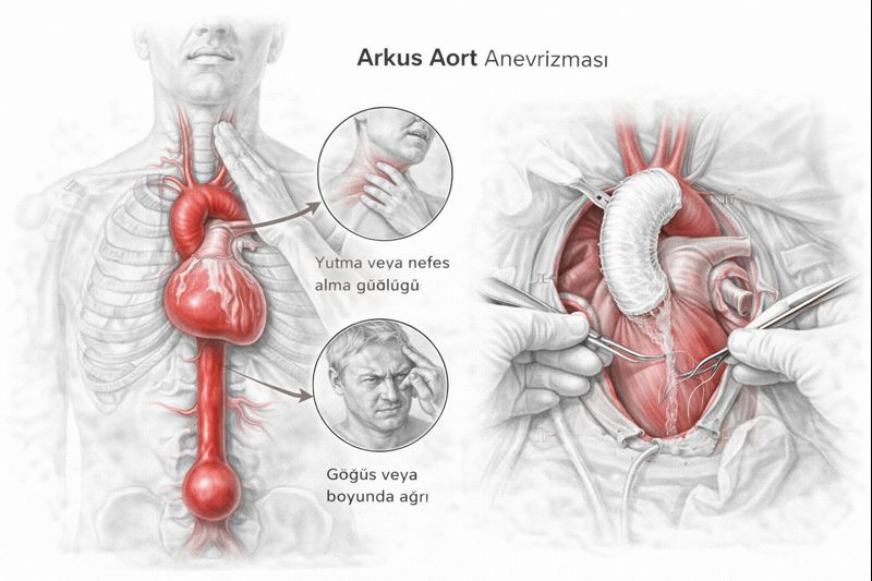
What Are the Symptoms?
Although aortic arch aneurysms generally progress silently, the following symptoms may be seen depending on the size:
- Dizziness and fainting
- Speech disorders or transient ischemic attacks (TIA)
- Arm and shoulder pain
- Chest pressure sensation
- Shortness of breath or cough
- Difficulty swallowing (if there’s pressure on the esophagus)
- Hoarseness
When there’s pressure on the arteries going to the brain, temporary or permanent neurological problems may occur. Therefore, these symptoms should be taken seriously.
How Is It Diagnosed?
Aortic arch aneurysm is usually detected during routine check-ups or during imaging related to another health issue. The diagnostic methods used are:
• CT Angiography (Computed Tomography Angiography)
It clearly shows the structure of the aortic arch and vessel diameter.
• MR Angiography
This radiation-free method is especially preferred in patients with chronic diseases.
• Transesophageal Echocardiography (TEE)
This ultrasound method performed by swallowing shows the arch region more closely.
Treatment of Aortic Arch Aneurysm
The treatment of aneurysms in this region is planned surgically. However, due to the location of the vessels and the necessity to protect the arteries going to the brain, it requires more specialized methods than standard surgical techniques.
1. Open Surgery
- The chest bone is opened.
- Circulation is maintained using a heart-lung machine.
- The aneurysmal segment is removed and replaced with an artificial vessel (graft).
- If necessary, the arteries going to the brain are redirected (bypass).
This operation is performed with advanced techniques such as hypothermia (lowering body temperature) and circulatory arrest (temporarily stopping circulation).
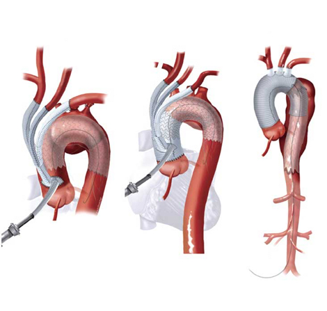
2. Hybrid Method
This method, which has become widespread in recent years, is a combination of open and closed surgery. It is preferred especially in high-risk patients. The process works as follows:
- The arteries going to the brain are redirected (bypass) without stopping the heart.
- Then, an endovascular graft (stent) is placed in the arch region, excluding the aneurysmal area.
These procedures are performed in hybrid operating rooms with specialized equipment.
Risks and Follow-up
The risk of rupture for an aortic arch aneurysm significantly increases when the vessel diameter exceeds 5.5 cm. An annual growth rate of more than 0.5 cm is also considered a surgical threshold.
During the follow-up process:
CT or MR angiography should be performed every 6-12 months.
Hypertension should be kept under control.
Smoking should be ceased, and blood lipids should be balanced.
Aortic arch aneurysm carries a high risk due to its location, which can directly affect the brain vessels. When diagnosed, treatment options should be individually planned by a cardiovascular surgeon. Thanks to advanced techniques such as hybrid surgery, it is now possible to achieve less invasive and more successful results.